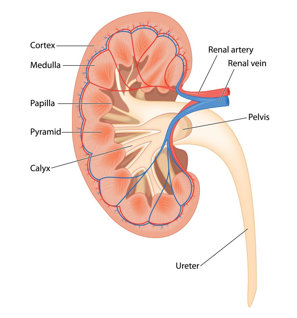
This image illustrates the internal anatomy of a kidney, highlighting its key structures and their functions. Each part of the kidney plays a crucial role in filtering blood, producing urine, and maintaining overall homeostasis in the body.
Cortex
The outermost layer of the kidney. It contains the glomeruli and the initial segments of the nephrons where blood filtration begins.
Medulla
- The inner region of the kidney, located beneath the cortex. It contains the renal pyramids and the loops of Henle, which are essential for concentrating urine.
Pyramid:
- Cone-shaped tissues within the medulla. Each pyramid contains tubules that transport urine from the cortical region to the calyces.
Papilla:
- The tip of each pyramid, which releases urine into the minor calyx.
Calyx (plural: Calyces):
- Cup-shaped structures that collect urine from the pyramids and channel it into the renal pelvis. There are minor calyces (smaller) and major calyces (larger).
Pelvis:
- A funnel-shaped cavity that collects urine from the calyces and channels it into the ureter.
Renal Artery:
- Supplies oxygen-rich blood to the kidney. It branches into smaller arteries within the kidney to reach the nephrons.
Renal Vein:
- Carries filtered blood away from the kidney back to the heart.
Ureter:
- A tube that transports urine from the renal pelvis to the bladder for storage until it is excreted.
The kidney’s intricate structure allows it to perform its vital functions of filtering waste products from the blood, regulating electrolytes, and maintaining fluid balance.
Quiz

Please note that our articles are not intended to guide personal health decisions.
This content has been curated by Renes Care. Unauthorized use or reproduction is prohibited.
© Renes Care. All rights reserved.